
Medical Design Record
Building a Mechanical Heart Valve
Follow the clinical, surgical and material advances that turned an experimental valve into durable mechanical life support.
Clinical problem
When the Native Valve Failed
Selected file: When the Native Valve Failed. Choose another file to update this evidence card.
Selected
Mechanical defect
Stenosis restricted forward flow, while regurgitation allowed blood to leak backward. Either defect could force the heart to work harder until compensation failed.
Major historical cause
Rheumatic heart disease left many mid-20th-century patients with scarred and deformed valves, particularly in the mitral position.
Unmet need
Early operations could separate fused leaflets or repair selected defects, but surgeons lacked a dependable substitute when a valve was too damaged to preserve.
The central challenge was not merely opening a valve, but replacing its one-way function continuously inside circulating blood.
Auxiliary prosthesis
Hufnagel’s Descending-Aorta Valve
Selected file: Hufnagel’s Descending-Aorta Valve. Choose another file to update this evidence card.
Selected
1952 implantation
Charles Hufnagel implanted a caged-ball prosthesis in the descending thoracic aorta of a patient with aortic regurgitation.
Location mattered
The device did not replace the diseased natural valve. It reduced part of the backward flow by acting farther downstream in the aorta.
Early construction
The valve used an acrylic chamber and ball. Later versions adopted a hollow nylon ball covered with silicone rubber to reduce weight and noise.
Proof supplied
Although physiologically limited, the implant showed that a synthetic moving valve could survive repeated cycling in the human circulation.
Hufnagel established implant feasibility, while leaving the harder task of replacing a valve inside the heart unresolved.
Surgical enabler
Cardiopulmonary Bypass Opens the Heart
Selected file: Cardiopulmonary Bypass Opens the Heart. Choose another file to update this evidence card.
Selected
Circulatory substitution
A heart-lung machine temporarily moved and oxygenated blood, giving surgeons time to open a bloodless heart and work on internal structures.
1953 milestone
John Gibbon used cardiopulmonary bypass successfully during closure of an atrial septal defect, demonstrating a practical route to intracardiac surgery.
New surgical possibility
With circulation supported outside the heart, surgeons could excise a damaged valve and secure a prosthesis at its natural annulus.
The valve mechanism and the heart-lung machine became complementary technologies: one enabled implantation, while the other sustained life afterward.
Orthotopic surgery
The 1960 Replacement Breakthroughs
Selected file: The 1960 Replacement Breakthroughs. Choose another file to update this evidence card.
Selected
Aortic replacement
In March 1960, Dwight Harken implanted a caged-ball prosthesis in the subcoronary aortic position, replacing the function of the removed natural valve.
Flexible mitral design
Nina Braunwald implanted a polyurethane-leaflet mitral prosthesis during the same period, demonstrating another approach to total valve substitution.
Long-term mitral success
On September 21, 1960, Albert Starr implanted the Starr–Edwards caged-ball valve in a patient who achieved sustained survival.
Credit distinction
These operations answered different “first” questions: aortic versus mitral position, design type, operative survival and durable clinical outcome.
Mechanical valve replacement emerged through several closely spaced operations, not a single uncontested moment of invention.
Clinical production design
Starr and Edwards Build for Durability
Selected file: Starr and Edwards Build for Durability. Choose another file to update this evidence card.
Selected
Shared roles
Surgeon Albert Starr defined the physiological and operative requirements. Hydraulic engineer M. Lowell Edwards developed and manufactured mechanisms that could meet them.
Design decision
After testing leaflet-like prototypes, the team adopted a free-moving ball that sealed against a circular seat and was retained by a cage.
Implant materials
Established models combined a silicone elastomer ball, a cobalt-alloy cage and a polyester sewing ring selected for durability and implant compatibility.
Iterative production
Clinical feedback drove repeated changes to cage geometry, ball composition, cloth coverage and manufacturing control during the early 1960s.
The Starr–Edwards valve converted a successful operation into a reproducible device that hospitals could implant in larger numbers.
Flow and material redesign
Tilting Discs and Pyrolytic Carbon
Selected file: Tilting Discs and Pyrolytic Carbon. Choose another file to update this evidence card.
Selected
Smaller occluder
Tilting-disc valves replaced the bulky ball with a pivoting circular disc, creating a lower-profile prosthesis and a less obstructed flow path.
Clinical introduction
The Björk–Shiley tilting-disc valve entered clinical use in 1969, followed by other single-disc designs with different pivots and opening angles.
Carbon transition
Jack Bokros and collaborators adapted pyrolytic carbon for blood-contacting valve components because it combined strength, wear resistance and favorable biocompatibility.
Unresolved surface problem
Improved carbon surfaces reduced several material limitations, but no mechanical geometry eliminated clot formation around artificial surfaces and pivots.
Disc geometry and pyrolytic carbon shifted valve engineering toward slimmer devices with better flow and extremely durable moving surfaces.
Modern mechanical pattern
The Bileaflet Valve Era
Selected file: The Bileaflet Valve Era. Choose another file to update this evidence card.
Selected
Two-leaflet opening
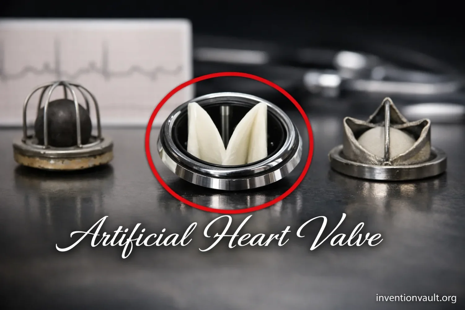
Two semicircular leaflets rotate apart to create one central and two lateral flow passages, distributing blood more evenly than a central ball.
1977 implant
The first surgical implantation of the all-carbon St. Jude Medical bileaflet valve took place in October 1977.
Design inheritance
Subsequent mechanical valves refined leaflet angles, hinge washing, housing contours, sewing cuffs and carbon processing without abandoning the bileaflet principle.
Persistent tradeoff
Modern mechanical valves offer exceptional structural durability, yet their blood-contacting surfaces still generally require long-term anticoagulation management.
Bileaflet construction became the dominant modern mechanical pattern because it balanced compact size, durability and improved forward flow.
The artificial heart valve was not invented in one event by one person. Charles Hufnagel proved in 1952 that a synthetic caged-ball valve could function in the human circulation, but his device sat in the descending aorta rather than replacing the damaged valve. True intracardiac replacement followed after cardiopulmonary bypass made open-heart surgery practical. In 1960, operations by Dwight Harken, Nina Braunwald and Albert Starr established different clinical milestones, while Starr’s partnership with engineer M. Lowell Edwards produced the first widely adopted, durable mechanical heart valve.
| Milestone | Date | People or team | What was established |
|---|---|---|---|
| Auxiliary caged-ball valve implanted | 1952 | Charles Hufnagel and clinical team | A mechanical one-way valve could operate in human circulation, although it did not replace the native valve. |
| Successful cardiopulmonary bypass operation | 1953 | John Gibbon and surgical team | Temporary extracorporeal circulation made controlled intracardiac surgery possible. |
| Orthotopic aortic valve replacement | March 1960 | Dwight Harken and team | A caged-ball prosthesis replaced valve function in the subcoronary aortic position. |
| Flexible mitral prosthesis implanted | March 1960 | Nina Braunwald and team | A polyurethane-leaflet prosthesis demonstrated a different route to mitral replacement. |
| Long-term successful mitral replacement | September 21, 1960 | Albert Starr, M. Lowell Edwards and team | The Starr–Edwards valve delivered sustained survival and became a reproducible clinical product. |
| Tilting-disc generation | 1969 onward | Viking Björk, Donald Shiley and other teams | A lower-profile disc reduced the central obstruction created by a ball. |
| All-carbon bileaflet valve implanted | October 1977 | St. Jude Medical development and surgical teams | Two pyrolytic-carbon leaflets established the dominant modern mechanical configuration. |
Dating the First
The 1952 Hufnagel operation was the first human implantation of an artificial valve, but the prosthesis was placed downstream from the heart. The 1960 procedures were the first replacements at natural valve positions. Long-term success, valve position and mechanism therefore produce different valid “first” dates.
Why a Replacement Valve Was So Difficult to Build
A natural heart valve is thin, flexible and responsive to very small pressure differences. It must open with little resistance, close rapidly without permitting substantial backflow and repeat this cycle tens of millions of times each year. Its surfaces are continuously exposed to blood, while its edges and supporting tissue absorb changing loads with every heartbeat.
A mechanical replacement did not need to resemble natural anatomy, but it had to reproduce the same one-way function. The moving element could not jam, fracture or wear rapidly. The housing had to remain anchored to living tissue, and every blood-contacting surface had to limit damage to blood cells and discourage clot formation.
The surgical problem was equally severe. Before dependable cardiopulmonary bypass, opening the heart long enough to remove a valve and sew in a substitute was not generally survivable. A practical artificial valve therefore depended on a system of innovations: extracorporeal circulation, improved anesthesia, blood management, sterile technique, sutures, implantable textiles and postoperative care.
Hufnagel’s Valve: A First Step Outside the Heart
Charles Hufnagel approached aortic regurgitation without opening the heart itself. In this condition, the aortic valve fails to seal properly, allowing blood to return toward the left ventricle after each contraction. Hufnagel placed his prosthesis in the descending thoracic aorta, where it could interrupt part of this reverse flow.
The device contained a freely moving ball inside an acrylic chamber. Forward blood flow pushed the ball away from its seat. When pressure reversed, the ball returned to the opening and sealed it. A cage retained the ball while allowing blood to pass around it.
This arrangement could not stop regurgitation into the coronary arteries or vessels supplying the upper body because those branches arose upstream from the implant. It also offered little help for a narrowed native aortic valve. Even so, the operation provided critical evidence: an artificial occluder could cycle in blood, remain mechanically contained and support a living patient.
Cardiopulmonary Bypass Changes the Surgical Boundary
The decisive enabling technology was the heart-lung machine. During cardiopulmonary bypass, venous blood is diverted to an external circuit, oxygenated and returned to the arterial circulation. The machine temporarily performs the pumping and gas-exchange work normally handled by the heart and lungs.
John Gibbon’s successful 1953 repair of an atrial septal defect showed that extracorporeal circulation could support a patient during an intracardiac operation. Other surgeons and engineers then refined pumps, oxygenators, tubing and operating methods. By the end of the decade, teams could stop and open the heart under controlled conditions, remove severely damaged valve tissue and attach a prosthesis to the remaining annulus.
This was the transition from adding an auxiliary valve elsewhere in the circulation to replacing the failed structure at its anatomical position.
The Multiple Valve-Replacement Firsts of 1960
Several teams reached important but distinct milestones in 1960. Dwight Harken performed an aortic valve replacement with a caged-ball device in March. The prosthesis was placed below the coronary openings, where it directly assumed the one-way function of the removed aortic valve.
Nina Braunwald implanted a flexible polyurethane mitral prosthesis during the same period. Her design used artificial leaflets and chord-like supports rather than a captive ball. It demonstrated that total mitral replacement was possible, although the materials and flexible construction of early leaflet prostheses presented durability challenges.
Albert Starr’s September operation with the Starr–Edwards valve supplied the landmark most often associated with successful mechanical mitral replacement. The patient survived long term, returning to an active life before dying years later from an unrelated accident. Starr and Edwards subsequently reported a series in which six of the first eight patients survived surgery.
Documented Clinical Milestone
The surviving clinical and institutional record supports September 21, 1960, as the first long-term successful implantation of a Starr–Edwards caged-ball valve in the mitral position.
How Starr and Edwards Turned a Prototype into a Product
M. Lowell Edwards initially wanted to develop a complete artificial heart. Starr, a cardiac surgeon at the University of Oregon Medical School, proposed beginning with a single valve. Their collaboration joined hydraulic engineering with operative observation: Edwards could rapidly build and alter mechanisms, while Starr could identify anatomical, flow and implantation problems.
The team initially explored designs that imitated natural leaflets. They eventually abandoned anatomical resemblance in favor of a mechanically simpler ball-and-cage system. The ball was unattached and could rotate, distributing contact and wear instead of repeatedly loading one hinge or edge.
The established Starr–Edwards models used a silicone elastomer ball retained by a cobalt-alloy cage. A polyester fabric sewing ring allowed the surgeon to anchor the valve to tissue. The cage geometry gave the ball enough travel to open the orifice while preventing its escape.
Clinical production required more than a working shape. Components needed consistent dimensions, smooth surfaces and predictable material properties. Early models were repeatedly modified in response to thrombosis, leakage, tissue overgrowth, blood-cell damage and wear. This feedback loop converted a hand-built experimental prosthesis into a controlled medical product.
How a Caged-Ball Mechanical Valve Works
- Forward pressure risesPressure on the inlet side exceeds pressure beyond the valve and pushes the ball away from its circular seat.
- The flow path opensThe cage limits ball travel while blood moves through the annular space between the displaced ball and valve housing.
- Pressure reversesAs the pumping chamber relaxes, downstream pressure becomes greater and drives the ball back toward the inlet.
- The seat closesThe ball contacts the circular seat and blocks reverse flow until the next pressure cycle begins.
The mechanism was robust, but the ball occupied the middle of the flow stream. Blood had to accelerate around it, creating regions of high velocity, turbulence and recirculation. The prosthesis was also relatively tall and heavy compared with later designs. These characteristics encouraged engineers to search for lower-profile occluders.
Why Materials Determined Clinical Survival
| Material or component | Historical use | Engineering value | Limitation or design concern |
|---|---|---|---|
| Polymethyl methacrylate | Early Hufnagel chambers and balls | Rigid, machinable and available for prototype production | Noise, bulk and long-term wear limited its suitability for later intracardiac designs. |
| Silicone elastomer | Ball occluders in Starr–Edwards valves | Lower density, resilience and quieter operation than a solid acrylic ball | Ball swelling, surface change and interaction with cage geometry required careful control. |
| Cobalt alloy | Cages and structural frames | High strength, corrosion resistance and experience in implanted orthopedic components | Blood-flow disturbances remained a geometric problem even when the metal itself was durable. |
| Polyester fabric | Sewing cuffs | Provided a flexible surface for suturing and tissue incorporation | Poor seating or excessive tissue growth could contribute to leakage or obstruction. |
| Pyrolytic carbon | Discs, leaflets and housings | Exceptional fatigue resistance, hardness and favorable blood compatibility | It reduces material-related problems but does not eliminate thrombosis or the need for anticoagulation. |
Pyrolytic carbon became especially important. Originally developed for high-temperature technical applications, it was adapted for valve components by materials engineer Jack Bokros and collaborators during the 1960s. Carbon coatings and structures offered the wear resistance required for billions of repeated cycles while producing a blood-contacting surface better suited to implantation than many early polymers and bare metals.
From Caged Balls to Tilting Discs
The next major design family replaced the central ball with a circular disc. In a tilting-disc valve, pressure rotates the disc around an offset pivot. The open disc divides the flow into a larger and a smaller passage rather than forcing all blood around a large spherical obstacle.
The Björk–Shiley valve, introduced clinically in 1969, became one of the best-known examples. Other designs, including the Lillehei–Kaster and Medtronic-Hall valves, altered the pivot, opening angle and housing. Their lower profile made implantation easier in restricted anatomical spaces and generally improved forward-flow performance.
The generation also demonstrated that small manufacturing details could have life-or-death consequences. Certain later Björk–Shiley convexo-concave models experienced outlet-strut fractures that could release the disc and cause sudden valve failure. The episode reinforced the need for fatigue testing, process control, traceability and long-term device surveillance.
Why the Bileaflet Design Became Dominant
A bileaflet valve uses two semicircular plates mounted in pivots within a circular housing. When open, the leaflets form three principal flow channels. When pressure reverses, both rotate closed against the housing.
The model developed for St. Jude Medical was the first all-pyrolytic-carbon bileaflet valve in clinical use, with its first surgical implantation recorded in October 1977. Its compact form and comparatively unobstructed opening provided better central flow than a caged ball. Pyrolytic-carbon leaflets and housing surfaces supplied the durability needed for lifelong cycling.
Later bileaflet valves refined the hinges because the pivot region is both mechanically essential and vulnerable to stagnant flow. Designers shaped the housing and leakage paths so small reverse jets could wash the hinge areas during closure. These controlled flows help clear blood from regions where clotting might otherwise begin, although they cannot make the device completely non-thrombogenic.
Mechanical Valve Versus Tissue Valve
Mechanical Valve
Manufactured from durable materials such as pyrolytic carbon and metal alloys. Structural life can be extremely long, but the artificial surfaces and hinges create clot risk that generally requires continuing anticoagulation management.
Tissue Valve
Constructed from treated animal tissue or human donor tissue. It usually avoids the same lifelong valve-related anticoagulation requirement, but biological leaflets can calcify, tear or stiffen and may eventually require another intervention.
These are not simply old and new versions of the same invention. They are different engineering compromises. Mechanical valves prioritize structural durability. Tissue valves more closely reproduce flexible leaflet motion and can simplify long-term medication requirements, but they have a finite risk of structural degeneration.
Anticoagulation Safety
Mechanical valves can develop obstructive clots or release emboli if anticoagulation is inadequate, while excessive anticoagulation increases bleeding risk. Starting, stopping or changing therapy requires individualized management by the patient’s clinical team.
What the Mechanical Valve Changed
Before dependable valve replacement, advanced valvular disease could progress beyond the reach of repair. A mechanical prosthesis gave surgeons a way to remove a structure that no longer opened or sealed and replace its essential function with a manufactured mechanism.
The achievement also altered medical-device engineering. Heart valves demanded fatigue resistance measured over billions of cycles, precise control of blood-contacting surfaces and close coordination between surgeons, materials specialists and manufacturers. Lessons from valve development influenced later work on blood pumps, ventricular-assist devices and other permanent cardiovascular implants.
Modern mechanical valves no longer resemble the tall acrylic device Hufnagel placed in the descending aorta. Yet they retain its basic engineering insight: pressure can move a captive occluder to produce passive one-way flow. The historical progression came from improving where that mechanism could be implanted, how much it obstructed blood, what materials could survive inside the body and how its unavoidable risks could be managed.
Questions People Ask About Artificial Heart Valves
Who invented the first artificial heart valve?
Charles Hufnagel developed and implanted the first artificial valve used in a human in 1952. Because it was placed in the descending aorta rather than at the diseased natural valve, Albert Starr and M. Lowell Edwards are often credited with the first widely successful intracardiac mechanical valve.
When was the first successful mechanical mitral valve implanted?
Albert Starr implanted the Starr–Edwards caged-ball valve in the mitral position on September 21, 1960. The operation is recognized as the first long-term successful mitral replacement with that prosthesis.
Was the Starr–Edwards valve the first caged-ball valve?
No. Hufnagel’s 1952 prosthesis already used a captive ball. The Starr–Edwards design adapted the principle into a smaller valve that could be sewn into the heart at the mitral and, in later models, aortic or tricuspid position.
Why did caged-ball valves fall out of routine use?
The central ball obstructed forward flow, produced less favorable flow patterns and required a relatively bulky cage. Tilting-disc and bileaflet valves offered lower profiles and improved hemodynamics.
What are modern mechanical heart valves made from?
Many modern designs use pyrolytic carbon for the leaflets and blood-contacting housing, combined with a fabric sewing cuff and selected metallic or carbon-based structural components.
Can a mechanical valve work without electricity?
Yes. It is a passive pressure-operated device. Changes in pressure across the valve move its ball, disc or leaflets. No motor, battery or electronic controller is required.
References Used for This Article
- The Caged-Ball Prosthesis 60 Years Later — historical review used for the Hufnagel, Harken and Starr–Edwards chronology, early materials and caged-ball design development.
- Smithsonian Institution, Starr–Edwards Heart Valve — museum record used for the prosthesis configuration, manufacturing history and early clinical significance.
- Oregon Health & Science University, Dr. Albert Starr — institutional history used for the Starr–Edwards partnership and long-term mitral implant account.
- Smithsonian Institution, St. Jude Medical Artificial Heart Valve — museum record used for the all-carbon bileaflet design and October 1977 implantation date.
- Smithsonian Institution, Bokros Carbomedics On-X Bileaflet Valve — used for the adaptation and continuing importance of pyrolytic carbon in mechanical valves.
- American College of Cardiology, Prosthetic Valve Guideline Summary — used for current mechanical-valve anticoagulation context.
- American Heart Association, Types of Replacement Heart Valves — used for the established durability and clot-risk distinction between mechanical and tissue valves.