| Item | Details |
|---|---|
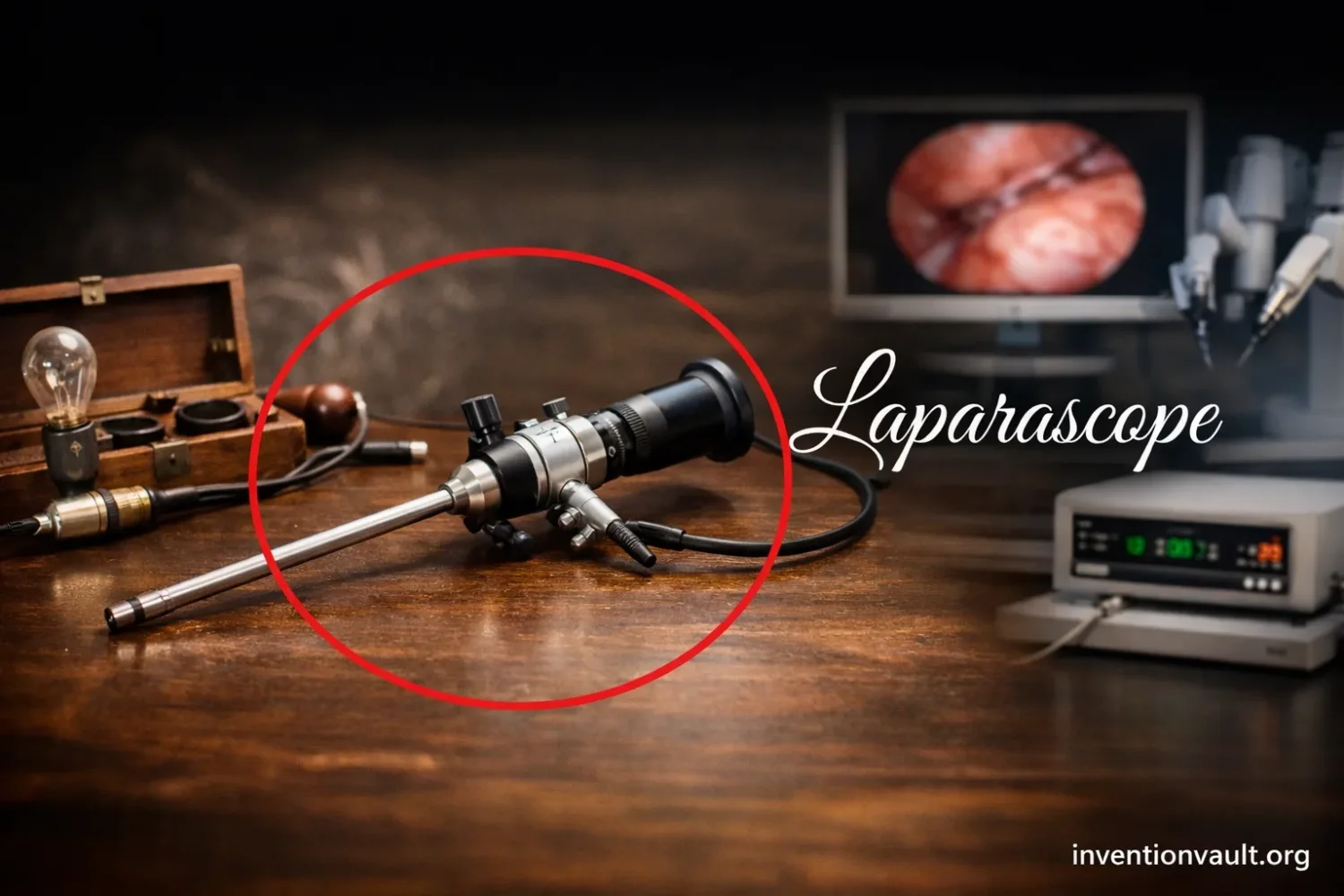
| What It Is | A laparoscope is a thin rigid instrument with a light and viewing optics used to examine, and later help operate within, the abdominal cavity through small openings. |
| Type of Invention | A cumulative medical instrument, not a one-day invention. The modern form appeared through staged improvements in optics, illumination, access, gas insufflation, image display, and surgical technique. |
| Single Inventor | No single inventor can fully claim the modern device. The earliest abdominal endoscopic step is tied to Georg Kelling, while the first human clinical use and publication are tied to Hans Christian Jacobaeus. Later pioneers made the instrument practical and widely usable. |
| Technical Roots | The laparoscope grew out of endoscopy, especially 19th-century work on illuminated scopes by figures such as Desormeaux and Nitze. |
| Earliest Abdominal Inspection | 1901: Kelling performed experimental abdominal inspection in a dog with a modified cystoscope after air insufflation, calling the method coelioscopy. |
| First Human Clinical Publication | 1910: Jacobaeus published his human clinical experience and helped establish the procedure in medical literature. |
| Early Naming | Historical labels included coelioscopy, laparothoracoscopy, organoscopy, culdoscopy, and later pelviscopy. |
| Important Access Advance | The move from plain air to CO2 insufflation and the later use of the Veres needle made access safer and the working space more controllable. |
| Important Optical Advance | Heinz Kalk improved viewing geometry; later the Hopkins rod-lens system and cold light sharply improved brightness and image quality. |
| Field That Carried It Forward | Gynecology played a central role, especially in fertility work, sterilization, and later egg retrieval for IVF. |
| Shift to Operative Use | The instrument became a true surgical platform only after better hemostasis, suturing methods, insufflators, and brighter optics were available. |
| Broad Surgical Takeoff | Late 1980s to early 1990s, when video laparoscopy and laparoscopic cholecystectomy pushed the method into general surgery. |
| Historical Importance | The invention changed abdominal surgery by pairing internal visualization with smaller incisions, shorter recovery, and a new model of surgical access. |
A fair history of the invention of the laparoscope starts with one correction: this was not a single flash of invention by one person holding one finished instrument. It was a layered technical achievement. Early endoscopists supplied the optical roots. Georg Kelling opened the abdominal chapter. Hans Christian Jacobaeus carried the method into human clinical practice. Later, gynecologists, physicists, and instrument makers turned an experimental viewing method into a dependable surgical tool. That longer story is what makes the laparoscope such an important invention in medical history.
The laparoscope did not arrive as a finished object. It became possible when optics, light, safe entry, gas control, and operative technique finally met.
What the Laparoscope Is
The word laparoscope refers to the instrument. Laparoscopy refers to the procedure performed with it. That distinction matters. Many articles blur the tool and the method, then rush straight to the late-20th-century rise of minimally invasive surgery. The history is older and more exact than that. The instrument began as a rigid viewing tube for the abdomen, then gained better lenses, brighter illumination, safer entry systems, and eventually camera-based image transmission. Only after those steps did it become the platform people now associate with modern minimally invasive surgery.
The First Step
Georg Kelling is usually placed at the beginning because he performed the first known abdominal endoscopic inspection in 1901. He used a modified cystoscope and air insufflation in a dog, and called the method coelioscopy.
The Clinical Turn
Hans Christian Jacobaeus is the figure most closely tied to human clinical laparoscopy. In 1910, he published his experiences in human patients and described both the promise and the hazards of the technique.
Who Invented the Laparoscope
If the question is framed strictly as “Who invented the laparoscope?”, the shortest accurate answer is this: no single person invented the modern laparoscope in one step. Kelling supplied the first abdominal endoscopic act. Jacobaeus established the human clinical procedure and documented it. Heinz Kalk improved the viewing system and made the tool more usable for diagnosis. Raoul Palmer, Hans Frangenheim, Kurt Semm, Harold Hopkins, and Karl Storz each solved a problem the early device could not solve well enough on its own.
That is why the laparoscope belongs to a class of inventions best understood as assembled inventions. The first version may open the door, but the invention reaches maturity only when later workers solve the practical obstacles. In the case of the laparoscope, those obstacles were plain: dim light, weak optics, blind access, poor control of abdominal distension, inadequate bleeding control, and limited ways to document or share what the operator saw.
The Endoscopic Roots Before 1901
The laparoscope did not appear from nowhere. It grew out of endoscopy. Nineteenth-century instrument makers and physicians developed illuminated scopes for body cavities that could already be reached more directly. Those devices were not yet laparoscopes, though they solved the first technical question: how to carry light and vision into the body. By the late 1800s, the work of Maximilian Nitze on rigid illuminated endoscopes gave later pioneers a tool they could adapt for the abdomen.
Georg Kelling and the 1901 Starting Point
Kelling’s place in history rests on more than a date. His work joined two lines of thought that had not yet been fully combined: endoscopic viewing and abdominal insufflation. He had been studying the effects of high-pressure abdominal air for the control of gastrointestinal bleeding. To observe what that insufflation did inside the abdomen, he introduced a cystoscope into the cavity of a dog. That synthesis is the real inventive leap. He was not just looking inside; he was creating an internal working space and then inspecting it.
Hans Christian Jacobaeus and the Human Clinical Record
Jacobaeus pushed the method into human medicine and into the literature that other physicians could read, judge, and reproduce. His 1910 publication described 17 laparoscopies and did something else that many short summaries omit: he also warned about the risk of organ injury during trocar insertion and recognized the need for training before clinical use. That matters historically. The invention was not just about seeing inside the abdomen. It was also about learning how to do so with discipline and repeatability.
How the Instrument Became Practical
The early instrument could demonstrate possibility. It could not yet support the wide clinical role that later generations would expect. The modern laparoscope emerged only when several technical problems were solved one by one.
| Problem | Advance | Why It Mattered |
|---|---|---|
| Poor viewing angle | Heinz Kalk’s improved lens geometry and forward-viewing design | Made inspection of abdominal organs more systematic and more useful for diagnosis. |
| Unsafe or awkward abdominal expansion | CO2 insufflation replaced plain air; later insufflation devices improved pressure control | Created a more stable and practical viewing space within the abdomen. |
| Difficult entry | Development and later adoption of the Veres needle | Helped establish pneumoperitoneum with a safer access method. |
| Heat and dim lighting | Cold light and later fiber-optic illumination | Improved visibility while reducing heat at the instrument tip. |
| Limited image clarity | Harold Hopkins’s rod-lens optics | Delivered brighter, sharper images and a wider field of view. |
| Little ability to operate | Hemostasis tools, loops, suturing methods, suction-irrigation, and safer insufflators | Turned laparoscopy from a mostly diagnostic method into an operative one. |
| Limited teaching and teamwork | Video display and camera systems in the late 20th century | Allowed the whole operating team to see the field and helped scale training. |
Heinz Kalk deserves more attention than he usually receives in popular retellings. He improved optics and developed a scope with a 135-degree lens system and a double trocar, helping make diagnostic laparoscopy more dependable. Later, the work of Harold Hopkins on rod-lens optics and the partnership with Karl Storz on better illumination gave the device a major leap in brightness and image quality. Without those improvements, the laparoscope would have remained interesting, but limited.
Why Gynecology Shaped the Invention’s History
Many short articles tell the history of the laparoscope as if general surgery carried the instrument from the start. That leaves out one of the most informative parts of the story. Gynecology was one of the main fields that kept laparoscopy alive, refined it, and gave it a broad clinical purpose long before the late-1980s expansion in general surgery.
Raoul Palmer advanced laparoscopic practice in fertility-related work, helped normalize abdominal positioning strategies such as the Trendelenburg position, and used laparoscopy in sterilization. Later, Patrick Steptoe helped codify laparoscopic sterilization and turned the method into something that could be taught more widely. That teaching role matters. An invention spreads only when it can be learned, not merely admired.
The same gynecologic line of development also fed directly into the early history of IVF. Laparoscopic egg retrieval by Steptoe and Robert Edwards formed part of the work that led to the birth of Louise Brown in 1978. That link is often missing from simplified histories, even though it shows how the laparoscope moved from diagnosis to a wider role in reproductive medicine and public awareness.
From Diagnostic Tool to Operative Platform
Early laparoscopy was mostly about seeing. Modern laparoscopy had to become about doing. That shift did not happen automatically. It required better methods for bleeding control, safer insufflation, improved instrument handling, and more precise optics. Figures such as Hans Frangenheim and Kurt Semm were central in this transition.
Semm is especially important because he treated laparoscopy as an operative system rather than a viewing curiosity. He developed an automatic CO2 insufflator, methods for coagulation and suturing, and tools that widened the surgical range of the instrument. His name is also tied to the first laparoscopic appendectomy. Some histories date that milestone to 1980, the year of the operation itself, while others date it to 1983, when the procedure entered published clinical discussion more clearly. That small date split is not trivial; it shows how invention history often turns on whether one counts the first performance or the first accepted report.
General surgery embraced laparoscopy more broadly only after video systems made the field easier to share on monitors. Once the operating team could see the same image, training sped up, teamwork changed, and the method moved faster into wider surgical practice. The late 1980s then became the period when laparoscopic cholecystectomy pushed the technique into everyday surgical debate and routine adoption.
Main Forms of Laparoscopic Practice
The laparoscope also has its own internal history of forms. The instrument stayed recognizable, but the way it was used widened over time.
- Diagnostic laparoscopy: used to inspect abdominal or pelvic organs and clarify disease or anatomy.
- Operative laparoscopy: used with working instruments to perform surgical tasks through small access points.
- Video laparoscopy: uses camera-based display rather than direct viewing through the eyepiece.
- Single-incision laparoscopy: reduces the number of access points, often through one main entry site.
- Hand-assisted laparoscopy: combines laparoscopic viewing with limited hand access for selected operations.
- Robotic-assisted laparoscopy: keeps the laparoscopic access model but adds robotic control and digital visualization.
These later forms do not replace the original invention story. They show how durable the original idea turned out to be. Once surgeons could see the abdominal cavity through small openings with enough light, enough space, and enough control, the instrument invited refinement rather than abandonment.
Why the Invention Matters in Medical History
The laparoscope changed more than instrument design. It changed how surgeons thought about access. In older abdominal surgery, the incision itself carried much of the burden of exposure. The laparoscope replaced part of that logic with internal visualization, controlled pneumoperitoneum, and a narrower route into the body. That was not a cosmetic shift. It altered surgical training, operating-room teamwork, postoperative recovery patterns, and the public image of abdominal surgery.
Seen that way, the invention of the laparoscope is not just a date in 1901 or 1910. It is a chain of linked advances that slowly turned a difficult experiment into a stable medical technology. Kelling and Jacobaeus stand at the head of the story. Kalk, Palmer, Frangenheim, Semm, Hopkins, Storz, and others explain why the story did not stop there.
References Used for This Article
- National Cancer Institute — Definition of Laparoscope: Official definition of the instrument and its core structure.
- PMC — The Development of Laparoscopy—A Historical Overview: Detailed timeline covering Kelling, Jacobaeus, Kalk, Palmer, Frangenheim, Semm, Hopkins, and later adoption.
- PubMed — Hans Christian Jacobaeus: Inventor of Human Laparoscopy and Thoracoscopy: Supports Jacobaeus’s role in early human clinical laparoscopy and his 1910 publication.
- PubMed — A Brief History of Endoscopy, Laparoscopy, and Laparoscopic Surgery: Useful for the transition from early abdominal inspection to late-20th-century video surgery.
- PubMed — Origin of Laparoscopy: Coincidence or Surgical Interdisciplinary Thought?: Explains how Kelling’s work joined insufflation research with endoscopic practice.
- Arizona State University — Laparoscopy: Summarizes early instrument changes, reproductive uses, and the later IVF link.
- Harvard Business School — Laparoscopy: Minimally Invasive Surgery: Helpful for adoption history, teaching, outpatient sterilization, and the IVF connection.
- PMC — The Evolution of Laparoscopy and the Revolution in Surgery in the Decade of the 1990s: Supports the role of optics, video systems, and late uptake in general surgery.